
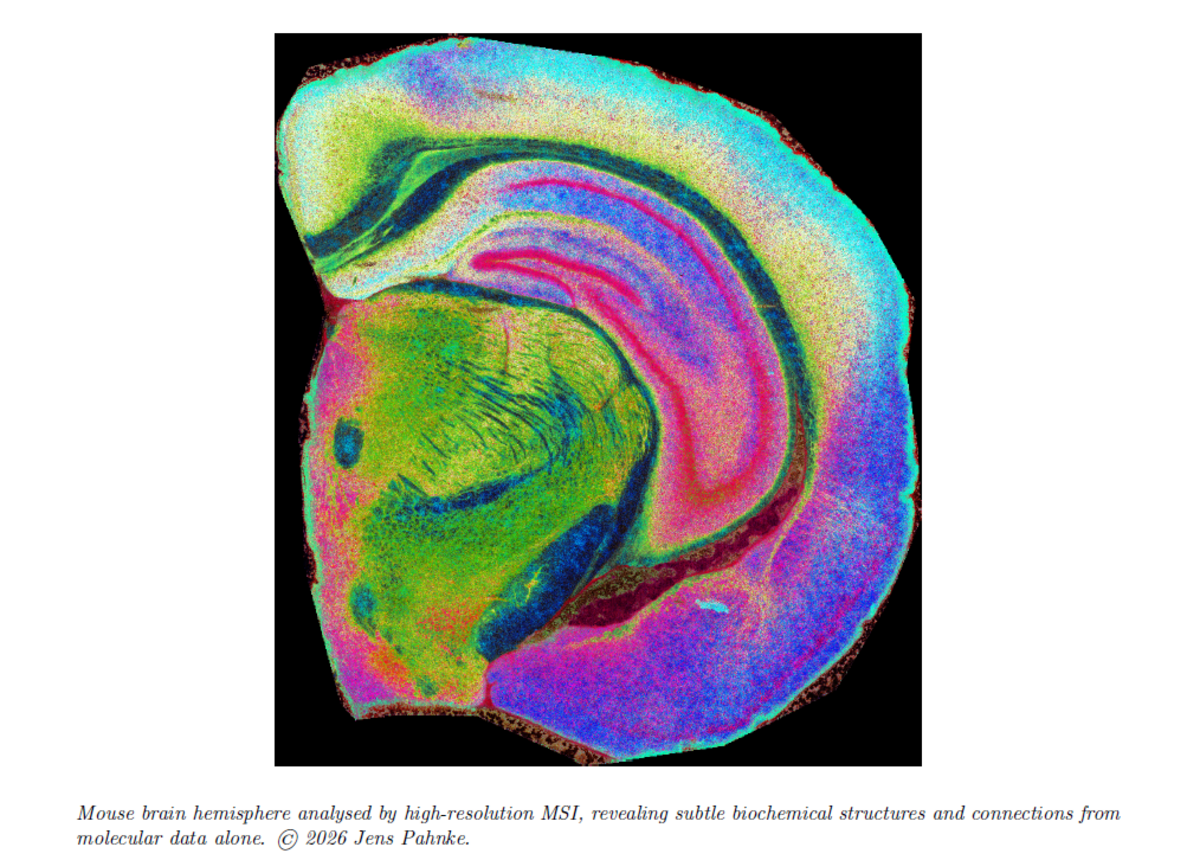
Mass spectrometry imaging measures thousands of molecular signals at defined positions in tissue, creating molecular “data cubes” that are rich but difficult to interpret. The publication of MSI-VISUAL in Science Advances places the work in a highly visible AAAS journal for major advances across science and underlines its relevance beyond one disease area or one technical community. The new MSI-VISUAL article addresses this bottleneck by providing visualization methods and interactive workflows that preserve tissue structure more faithfully. Researchers and pathologists can select regions of interest, compare molecular profiles, and map specific mass-to-charge signals back into the tissue image. The method can reveal subtle brain structures, disease-related changes, single large nerve cells, inflammation, and organ damage using molecular signals alone.
The impact reaches far beyond dementia research. In cancer pathology, for example, diagnosis increasingly depends on recognizing molecular heterogeneity: tumor borders, invasive fronts, resistant subclones, necrotic zones, immune-rich regions, and metastases may differ chemically before they differ morphologically. MSI-VISUAL is designed to expose precisely these hidden molecular patterns. It can therefore become a bridge between classical pathology and spatial molecular diagnostics, supporting future workflows in which tissue sections are not only stained, but chemically read. For patients, this could support earlier diagnosis, better patient stratification, and faster evaluation of whether new treatments are working.
MSI-ATLAS demonstrates what becomes possible when such visualization is combined with expert annotation and explainable machine learning. Using MSI data alone, the team generated a detailed computational atlas of the mouse brain, covering 123 region types and 191 polygonal annotations. The atlas shows that brain regions have characteristic lipid and metabolite signatures. Importantly, regions that are anatomically or functionally connected can share lipid patterns, suggesting that lipid composition reflects brain networks, not only local cell composition.
This is highly relevant for Alzheimer’s disease and other dementias. APOE is the strongest common genetic risk factor for late-onset Alzheimer’s disease, and ABCA7 is another major risk gene. Both are linked to lipid transport and lipid handling. MSI-ATLAS provides a spatial framework for understanding why these proteins matter: if brain regions and networks depend on precise lipid organization, then disturbed lipid transport may alter network vulnerability, amyloid plaque chemistry, and disease spread. The atlas also highlights plaque-associated lipid signals, including ganglioside-related patterns, and offers testable hypotheses about how pathological deposits relate to surrounding brain structures. It may also help distinguish overlapping disease processes in the same brain, for example Alzheimer’s pathology versus vessel-related damage.
The scientific importance is therefore twofold. First, the studies deliver practical tools for future diagnostics and discovery-oriented pathology. Second, they provide a new conceptual view of the brain as a molecularly organized lipid landscape. For dementia research, this supports a shift from studying single molecules in isolation toward understanding spatial lipid networks, transport mechanisms, and region-specific vulnerability. For biomedical research more broadly, the same approach can be applied to metabolites, lipids, peptides, tumors, inflammatory lesions, kidney disease, and other complex tissues.
Author information
Jacob Gildenblat is a Dr.-Ing. candidate in the Pahnke Lab under the supervision of Prof. Jens Pahnke. His work focuses on computational methods, visualization, and machine learning for mass spectrometry imaging.
Prof. Dr. med. Dr. rer. nat. Jens Pahnke, E.F.N. leads the Translational Neurodegeneration Research and Neuropathology Lab at the University of Oslo and Oslo University Hospital, with affiliated professorships and collaborations in Lübeck, Riga, and Tel Aviv. His laboratory focuses on translational research into neurodegenerative
diseases, especially dementias and movement disorders. He has received several research prizes, including the Alzheimer Research Initiative Prize 2008, the Oswald Schmiedeberg Prize 2022, and the Norwegian Dementia Research Prize 2025. His work is supported by the Norwegian Research Council, the South-Eastern Norway Regional Health Authority, and the EU EIC Pathfinder program. Homepage: https://www.pahnkelab.eu
Contact: Prof. Jens Pahnke, Pahnke Lab, University of Oslo / Oslo University Hospital, jens.pahnke@medisin.uio.no
Publications
MSI-VISUAL: Gildenblat, J. & Pahnke, J. Truthful visualizations for mass spectrometry imaging enable high spatial resolution interactive m/z mapping and exploration. Science Advances. https://doi.org/10.1126/sciadv.aed3650
MSI-ATLAS: Gildenblat, J., Stamnæs, J. & Pahnke, J. Mass spectrometry imaging-based explainable machine learning reveals the biochemical landscapes of the mouse brain. Free Neuropathology 7, 9 (2026). https://doi.org/10.17879/freeneuropathology-2026-9413
